When your doctor suspects a heart problem, they don’t just listen with a stethoscope. They look inside. Two tools do most of that looking: cardiac MRI and echocardiography. Both show your heart’s structure and function, but they’re not the same. One is fast, cheap, and everywhere. The other is precise, detailed, and not always easy to get. Knowing the difference helps you understand why one test is ordered over the other-and what each can really tell you about your heart.
How Echocardiography Works: The Sound Wave View
Echocardiography uses sound. High-frequency ultrasound waves bounce off your heart, and the echoes turn into moving pictures on a screen. It’s been around since the 1950s, and today, it’s the most common heart test in the world. About 15 million echocardiograms are done in the U.S. every year.
Why so popular? It’s portable. You can roll the machine into an ER, a hospital room, or even a doctor’s office. No prep. No waiting. Results come in minutes. A typical echo takes 20 to 45 minutes. It shows how well your heart pumps, the size of its chambers, and whether valves leak or narrow.
Normal values? Left ventricular ejection fraction (LVEF) between 50% and 75% means your heart is squeezing strongly. The wall thickness of the septum (the wall between the two lower chambers) should be between 6 and 11 mm. The left ventricle’s width at rest (LVEDD) is usually 37 to 56 mm. These numbers are measured using formulas that assume your heart is shaped like a football or ellipse. That’s a big assumption-and it’s where echo starts to stumble.
How Cardiac MRI Works: The High-Definition Scan
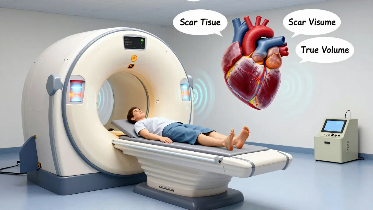
Cardiac MRI uses magnets and radio waves, not sound. You lie inside a tube-like machine that creates a powerful magnetic field (usually 1.5 or 3 Tesla). It takes 30 to 60 minutes. No radiation. No needles-unless contrast dye is needed.
What makes MRI special? It doesn’t guess. It measures. While echo uses math to estimate heart volume, MRI scans the whole heart in 3D, slice by slice. No assumptions. No rounding. The numbers are direct. For example, normal left ventricular volume in men is 67 to 155 mL. In women, it’s 55 to 105 mL. LV mass? 49 to 115 g for men, 37 to 81 g for women. These aren’t estimates. They’re actual measurements.
But MRI’s real superpower is tissue. It can see scar tissue, inflammation, or fat deposits inside the heart muscle. Late gadolinium enhancement (LGE) highlights areas of fibrosis-scar from past heart attacks or chronic disease. That’s something echo can’t do at all.
Accuracy: When Echo Lies and MRI Doesn’t
Here’s the hard truth: echocardiography often gets the numbers wrong. A 2011 study in the Journal of Cardiovascular Magnetic Resonance found echo consistently underreported heart chamber sizes by nearly 100 mL and overestimated wall thickness by more than 1 mm. Why? Because it relies on geometric models. If your heart doesn’t look like a perfect oval, the math breaks.
Cardiac MRI? It’s the gold standard. The European Society of Cardiology says it’s the reference method for measuring heart volume and muscle mass. Why? Because its inter-observer variability is just 2.6%. That means if two different radiologists look at the same MRI, they’ll agree within 2.6%. Echo? That number jumps to 6.8%. In a 2023 study published in JACC: CardioOncology, echo underestimated LVEF by a median of 3% compared to MRI. That might sound small, but in cancer patients being monitored for heart damage from chemo, that 3% can mean the difference between catching early damage or missing it entirely.
One study found that 10% of oncology patients were misclassified for heart risk because echo gave a false reading. MRI caught it.
When Each Test Is Used
Most people start with echo. It’s the first step. If you have shortness of breath, chest pain, or a heart murmur, echo is the go-to. It’s quick, safe, and tells you 80% of what you need to know.
But when echo gives unclear answers? That’s when MRI steps in.
- Unclear results: If your echo images are blurry because you’re overweight or have lung disease, MRI fills the gap.
- Heart muscle diseases: Hypertrophic cardiomyopathy, arrhythmogenic right ventricular dysplasia, or infiltrative diseases like amyloidosis? MRI sees the tissue changes before function drops.
- Scar detection: After a heart attack, MRI shows exactly where the scar is. Echo can’t.
- Pre-surgery planning: For complex valve repairs or congenital heart defects, MRI gives surgeons a 3D map.
- Monitoring treatment: In heart failure or after chemotherapy, MRI tracks changes more reliably than echo.
Cardiologists use echo daily. But they turn to MRI when they need certainty. One study of 127 cardiologists found 76% used MRI only after echo failed to give clear answers.
The Downside of MRI: Access and Cost
Cardiac MRI isn’t perfect. It’s expensive. A single scan costs $1,500 to $3,500. Echo? $500 to $1,500. That’s why echo is still the workhorse.
And then there’s access. In 2023, a survey by the American College of Cardiology found 68% of patients waited over two weeks for a non-urgent cardiac MRI. Community hospitals? Only 35% offer same-week MRI. Most rely on echo.
Not everyone can have an MRI. If you have a pacemaker, defibrillator, or certain metal implants, you’re often excluded. Even with new low-field MRI machines (like Siemens’ 0.55T system introduced in 2023), some devices still pose risks. Arrhythmias like atrial fibrillation can also mess up the images because the heart isn’t beating regularly.
Echo doesn’t have these limits. It works on anyone. Even in the middle of the night, in the ER, with a crashing patient, echo is there.
Training and Expertise
Becoming skilled at echo takes about 300 to 500 supervised scans. That’s 6 to 12 months of practice. Most sonographers hit proficiency within a year.
Cardiac MRI? It takes 1,000 to 1,500 interpreted studies. That’s 18 to 24 months of training. Fewer people are certified in MRI, which adds to the wait times.
Reporting is another difference. Echo systems have built-in templates that auto-fill measurements. MRI requires specialized software like cvi42. Radiologists need extra training to use it. That’s why many community hospitals outsource MRI reads to academic centers.
What’s Changing? The Future of Heart Imaging
Technology is closing the gap. Philips’ EPIQ CVx system, released in March 2023, uses AI to auto-calculate LVEF and wall motion. It cut inter-observer variability down to 4.2%-much closer to MRI’s numbers.
On the MRI side, 0.55T scanners are making it possible to scan patients with older pacemakers. New techniques like T1 and T2 mapping give doctors numbers for tissue health-like measuring water content in muscle-without needing contrast dye.
Experts predict hybrid protocols will be common by 2030. Start with echo. If something looks off, jump straight to MRI. No more guessing. No more delays. Just clear answers.
For now, echo is the first look. MRI is the deep dive. You don’t need both. But knowing which one your doctor chose-and why-helps you understand your heart’s story.
Is cardiac MRI better than echocardiography?
Cardiac MRI is more accurate for measuring heart size, muscle mass, and detecting scar tissue. It’s the gold standard for detailed assessment. But echocardiography is faster, cheaper, and works for most routine checks. Neither is "better" overall-they serve different purposes.
Can you have both tests on the same day?
Yes, but it’s rare. Echo takes 30 minutes. MRI takes 45 to 60. Most doctors space them out unless there’s an urgent reason. If you’re being evaluated for a complex condition like myocarditis or cardiomyopathy, your care team might schedule both within a week.
Why does my doctor order an MRI after an echo?
Echo often gives a good first look, but it can be unclear if you’re overweight, have lung disease, or if the heart looks abnormal. MRI gives a clearer picture of size, shape, and tissue health. If echo says "maybe" or "inconclusive," MRI gives the answer.
Is cardiac MRI safe if I have a pacemaker?
It depends. Older pacemakers and defibrillators are still a strict no for MRI. Newer models are labeled "MRI-conditional," meaning they’re safe under specific conditions. Always tell your doctor about any implants. A 0.55T MRI machine, introduced in 2023, is safer for some patients with older devices, but not all.
How long does it take to get results from a cardiac MRI?
The scan itself takes 30 to 60 minutes. But the analysis is complex. A radiologist needs time to review hundreds of images and use specialized software. Results usually take 3 to 7 days. In urgent cases, your doctor may get a preliminary read the same day.
Does echocardiography detect heart scarring?
No. Standard echocardiography can’t detect scar tissue or fibrosis in the heart muscle. That’s one of the main reasons cardiac MRI is used-it shows areas of scar using late gadolinium enhancement. If your echo shows reduced function but no clear cause, MRI may be ordered to check for hidden scarring.
What to Do Next
If you’ve had an echo and your doctor says "follow up with MRI," don’t panic. It doesn’t mean you have a serious problem. It just means they need more detail. If you’re waiting weeks for an MRI, ask if your clinic has a priority pathway for cardiac cases. Some centers fast-track patients with symptoms like unexplained shortness of breath or abnormal EKGs.
If you’re scheduled for an MRI and you’re nervous-about the noise, the space, or the contrast dye-talk to the technologist. They’ve seen it all. They can explain what to expect. And if you can’t have MRI? Don’t worry. High-quality 3D echo, especially with AI tools, is getting closer to MRI’s accuracy every year.
Your heart doesn’t need perfect imaging. It needs the right test, at the right time. Whether it’s sound waves or magnets, the goal is the same: to see what’s happening inside-and act before it’s too late.
phyllis bourassa
I love how echo is still the go-to for most docs... but let's be real, if your heart looks like a squished potato on ultrasound, you're getting an MRI next week. I had mine after my echo said 'maybe hypertrophy' and my cardiologist just sighed and said 'yeah, we'll need to see the real deal.' Took 3 months to get in. Worth every second.
Patrick Jackson
MRI is like seeing your heart in 4K HDR... echo is like watching it through a foggy window with a flashlight. 😅 I work in oncology and saw a patient get chemo for 6 months because echo said her EF was 52%... MRI showed 41%. She had silent damage. We almost lost her. Tech is amazing but we still rely on flawed tools. Sad but true.
Pranay Roy
You know who really benefits from MRI? The insurance companies. They delay it for months so they can deny coverage later. I’ve seen it. Echo is cheaper so they push it. But the real reason? Hospitals don’t want to pay for MRI techs or maintenance. It’s all about profit. Not science.
Joe Prism
Echo: fast. MRI: true. Doctors pick based on what they can afford. Not what’s best. Simple as that.
Andrew Poulin
If your echo is blurry you're not fat you're just unlucky. Get the MRI. Stop wasting time. Your heart doesn't care about your insurance deductible.
Weston Potgieter
Echo is basically guesswork with a camera. MRI is truth with magnets. Why do we still use echo? Because the system hates precision. It loves ambiguity. It loves delays. It loves paperwork. And it loves making you wait 6 weeks for a scan that could’ve been done in 2 days if you lived in a city with real healthcare.
Vikas Verma
LVEF estimation error of 3% may seem minor but in high-risk cohorts it translates to significant clinical misclassification. MRI-based quantification remains the reference standard per ESC guidelines. Adoption of AI-enhanced echo is promising but not yet sufficient to replace gold standard imaging in complex cardiomyopathies.
amber carrillo
I just got my MRI after my echo came back 'borderline.' I was terrified. But the tech was so kind. She explained every step. And when they found the tiny scar? It wasn't a disaster. Just something to watch. I’m so glad I didn’t panic. You’re not alone.
Tim Hnatko
I’ve read every word. Echo is the first look. MRI is the deep dive. Neither is perfect. But together? They save lives.
Joey Pearson
If your doctor says MRI after echo, don’t stress. It’s not a death sentence. It’s a clearer picture. And honestly? I’d rather know the truth than live in the dark. My mom’s heart scare? MRI caught it early. We got ahead of it. You’ve got this.